
UA researcher's 3-D tumor models could improve cancer treatment
The National Institutes of Health (NIH) is supporting the work of a University of Akron researcher who may hold the key to improving the effectiveness of cancer treatments. The agency has awarded Hossein Tavana a two-year, $511,000 grant to fund his ongoing efforts to improve the testing and effectiveness of anticancer drugs.

Dr. Hossein Tavana (center), is assisted in his research by graduate students Ehsan Atefi (left) and Stephanie Lemmo (right).
Tavana, an assistant professor of biomedical engineering in the College of Engineering, has developed a method to generate 3-D cultures of cancer cells (spheroids) that better model tumors in the body. These improved models have the potential to dramatically improve the screening and discovery of effective chemotherapeutics, Tavana says.
In support of this novel and promising technology, the NIH awarded Tavana its R21 grant, which is defined by the NIH as a developmental research grant intended to support "exploratory, novel studies that break new ground or extend previous discoveries toward new directions or applications."
3-D better mimics cancer cells in body
In contrast to the traditional 2-D culture of cancer cells, in which a thin layer of cells is treated on a flat, plastic dish, a 3-D culture of solid, clustered cells allows researchers to test chemotherapeutic drugs on tumor models that better mimic the complex environment of cancer cells in the body. This method makes drug screening and discovery more efficient and cost-effective, Tavana explains.
He points out that pharmaceutical companies spend an enormous amount of time and money on testing thousands of compounds on 2-D "monolayer cultures" without being able to adequately predict how the drugs will behave in the 3-D environment of the body.
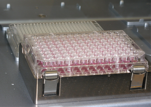
A 384-well plate containing culture media. Cancer cells are immersed in the media, where they aggregate into spheroids.
"In a 2-D culture, a small concentration of a drug can kill all of the cells," Tavana says. "But that same drug might only penetrate the peripheral cells, and not the core, of a solid 3-D tumor in the body. So, if you are able to conduct those initial tests with a three-dimensional model that is a closer mimic of the actual tumor, then you would save a lot of time and money.
Testing on 3-D models would allow researchers to determine with greater accuracy which drugs will best treat particular forms of cancer, Tavana says, eliminating the need to treat patients with a battery of drugs in the hope of finding something that works.
Personalized treatment
"So, if this is successful, this may be the next step toward personalized medicine where a patient's own cells are tested using this technology," Tavana adds. "Rather than throwing a bunch of different drugs into a patient's body, we can say, 'This particular patient, based on this test, will most likely benefit from this chemo drug.'
"We are at the cutting edge of the research going on in 3-D culturing of cancer cells," he says, citing Johns Hopkins University and Cornell University as two of the few sites doing comparable research in 3-D culturing.
But UA's advantage, Tavana says, is its novel method of generating 384 cell spheroids, or aggregates, "robotically and in a single step," rather than just one aggregate at a time. This will significantly expedite the efficiency of drug testing, he adds.

Graduate student Stephanie Lemmo manually loads the well plate. In practice, this is done by the robotic liquid handler.
"One thing that got the reviewers [from NIH] excited is that we are able to use a robot," Tavana says. "One of the reasons drug companies are not moving toward using three-dimensional cultures is that existing 3-D culturing methods are manual and hard to perform."
The robot, equipped with rows of pipettes, dispenses cancer cells into 384 small wells, or miniature test tubes, each of which contains a liquid that provides nutrients to the immersed cells, allowing them to aggregate as they would in the body, resulting in 384 physiologic tumor models.
"We are very excited that our research has been recognized by the NIH," says Tavana, who last month received a separate, three-year, $310,000 grant from the National Science Foundation for his research on the microenvironment of stem cells.
Collaboration leads to results
"This R21 grant will help fully develop and validate our technology and catalyze our ongoing efforts in developing translational cancer technologies," he says. "And of course, this award is a product of hard work by two of my [graduate] students, Ehsan Atefi and Stephanie Lemmo, and several years of collaboration with Dr. Gary Luker from the University of Michigan Medical School.”
Once Tavana and Luker, associate professor of microbiology and immunology at the University of Michigan, validate their technology by showing consistent generation of 3-D tumor models, they will apply for an R33 grant, which supports further, more substantial biological validation of the technology.
Tavana and his research team are currently testing cell lines — cells that have been made immortal so they can be reused over and over — of triple negative breast cancer and skin cancer.
"Currently, breast cancer [Luker's area of expertise] has the highest rate of mortality among women, so there is a pressing need to move forward," Tavana says.
Next year, during the second phase of their validation, Tavana and his team will use primary, patient-derived cells, which will allow them to test drugs under more realistic conditions.
"Cell lines are easy to handle and maintain," Tavana says, "but over time they lose some of their physiologic characteristics due to frequent passaging in culture dishes. The advantage of using primary cells is that these cells have all their original characteristics and functions, and we will be able to elicit more physiologic response from them when treated with drug compounds."
![]() Story by Nicholas Nussen
Story by Nicholas Nussen
Media contact: Denise Henry, 330-972-6477 or henryd@uakron.edu.