
Multiphoton Laser Scanning Microscope
Rm 210 NPIC
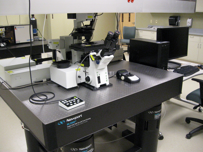
Olympus Fluoview 1000 MPE Multiphoton Laser Scanning Microscope
Background
Multiphoton fluorescence microscopy is a powerful research tool that combines the advanced optical techniques of laser scanning microscopy with long wavelength multiphoton fluorescence excitation. At high photon densities, two (or more) photons can be simultaneously absorbed by combining their energies to provoke the electronic absorption of a fluorophore in the specimen. This will then result in fluorescence emission in the visible region. Excitation in multiphoton microscopy occurs only at the focal point of a diffraction-limited microscope objective, thus providing the ability to optically section thick biological and other transparent specimens in order to obtain three-dimensional resolution.
Because the energy of a photon is inversely proportional to its wavelength, the two photons should have wavelengths about twice that required for single-photon excitation and the excitation sources need to be emitting in the red or infrared regions. In addition, unlike the case for single-photon absorption, the probability that a given fluorophore will simultaneously absorb two photons is a function of both the spatial and temporal overlap between the incident photons. In fact, photon concentration must be approximately a million times that required for an equivalent number of single-photon absorptions. This is accomplished with high-power mode-locked pulsed lasers, which generate a significant amount of power during pulse peaks.
Individual optical sections are acquired by raster scanning the specimen in the x-y plane with the laser beam and detecting the induced fluorescence, and a full three-dimensional image is composed by serially scanning the specimen at sequential z positions. Because the position of the focal point can be accurately determined and controlled, multiphoton fluorescence is useful for probing selected regions beneath the specimen surface. The highly localized excitation energy minimizes photobleaching of fluorophores attached to the specimen and reduces photodamage.
In multiphoton microscopy, photons emitted through fluorescence originate almost exclusively from the objective focal plane, eliminating the requirement for descanned detection and permitting more flexible detection geometries. This increased versatility leads to a considerable improvement in fluorescence detection efficiency.
This text is an adaptation from http://www.olympusmicro.com/primer/techniques/fluorescence/multiphoton/multiphotonintro.html
Applications
Olympus' Fluoview FV1000-MPE, is a multiphoton laser scanning microscope that allows fluorescence imaging deep within specimens. Utilizing ultrashort pulsed IR laser, the Olympus Fluoview MPE is able to image hundreds of microns into a specimen thanks to the penetration of IR light and uniquely designed long working distance objectives. The Fluoview-MPE provides cutting-edge technology for various areas of scientific research such as neuroscience and cell biology. Optimal for in-vivo observation requiring deeper imaging.
Specifications
- Laser
- Mode-locked Ti:sapphire laser: a femtosecond laser equipped with a group velocity dispersion correction/control device, laser power unit, water-cooled circulating chiller
- Model MaiTai DeepSee-OL: 710 nm — 1040 nm
- Scanning unit
- Scanning method: Light deflection via 2 galvanometer scanning mirrors
- Scanning modes: Pixel size: 64 x 64 — 4096 x 4096 pixels
- Scanning speed (pixel time): 2 μs — 200 μs; High-speed scanning mode: 16 frames/s (256 x 256)
- Dimensions: Time, Z, (wavelength) (or any combination thereof)
- Line scan: straight line (includes rotation), free line, point
- Detector for multiphoton imaging:
- Photomultiplier (PMT) (4 channels), fluorescence wavelength can be selected with the dedicated filter cube
- The high sensitivity PMT installed near the objective lens eliminates the need to use confocal aperture and makes possible detection of even scattered fluorescence
- IR excitation light is removed by high fluorescence transmission IR cut filter, this provides fluorescent imaging with high S/N ratio
- Optics with the infrared laser for multiphoton imaging
- Integrates a multiphoton near-infrared pulsed laser in the scanning unit
- High speed shutter system with acousto-optical modulator (AOM) – reduces damage to specimen during observation
- Continuously variable output using AOM (1 – 100%, 0.1% increments)
- Inverted microscope Olympus IX81
- Z-drive
- A motorized focus module inside the microscope is used
- Minimum increment: 0.01μm
- Water immersion objective designed specifically for multiphoton imaging: ULTRA 25x, NA 1.05
- Two-photon dedicated optical design for optimal performance in the NIR range
- 82% transmittance from 400nm to 1000nm
- Deep imaging correction collar creates a very tight focusing spot, ideal for multiphoton excitation
- Fluorescence signal can be detected through the full 1.05 NA, filling the entire back aperture and improving fluorescence detection efficiency
- Objective design offers excellent compensation for either cover slip or non-cover slip applications