
X-Ray Photoelectron Spectrometer (XPS)
Rm 212 NPIC
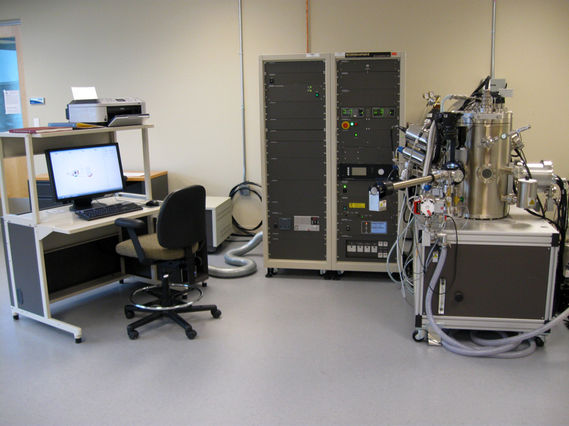
PHI VersaProbe II Scanning XPS Microprobe
Background
X-Ray Photoelectron Spectroscopy (XPS) is one of the most extensively used analytical techniques due to its ability to analyze with high sensitivity the elemental composition and chemical bonding in the top 10 nm near the surface of the specimen. Modern XPS systems are sophisticated and possess a high level of automation. The analysis is performed in ultra-high vacuum that needs to be maintained 24/7. Nevertheless state-of- the-art systems like the one that is installed at the SOA Center are characterized with user friendly operation, high reliability of the collected data, ease of chemical interpretation through specialized software. XPS measures the kinetic energies of core photoelectrons that originate from specimen atoms and leave the specimen when it is irradiated by a focused beam of soft X-ray photons (1 – 2 keV). Part of the x-ray beam energy is used to compensate for the characteristic binding energy of the core electrons that keeps them bound to the nucleus. Therefore recording the kinetic energy and and knowing the energy of the X-ray photons is enough for determination of the binding energies of electrons in all elements. XPS gives unique spectral signatures for each element and can be used for their most accurate identification.
XPS has the following unique analytical features:
- Provides information of the binding energy of core electrons in atoms – X-ray Photoelectron Spectra. Analysis needs ultra-high vacuum (UHV)
- Intrinsically surface specific technique – signal from the top 10 nm of the sample
- Provides elemental information for all elements except H and He (solid samples only). Conductive and insulating materials. Nondestructive technique
- Provides information for chemical bonding in specific molecular configurations through measurement of chemical shifts
- Quantitative analysis – relative atomic % concentration can be determined, sensitivity more than 0.1%.
Applications
Polymers, metals, ceramics, catalysts, corrosion, biomaterials, adhesion, semiconductors, dielectric materials electronics packaging, magnetic media, thin film coatings
Specifications
- Scanned, micro-focused, monochromatic (Al Kα) X-ray beam (E = 1486 eV), 8 to 100 um diameter beam spot
- X-ray beam induced secondary electron imaging (SXI) for surface topology review and selection of regions/points of interest for XPS analysis
- Automated dual beam charge neutralization to help analysis of insulating samples
- Automatic sample alignment for optimal signal collection
- Point-and-Click analysis area selection
- Single and Multi-point analysis
- 128 channel detection. Energy resolution 0.1 eV
- High sensitivity large area XPS (1400 x 100 um)
- High sensitivity micro area XPS
- Chemical state imaging and elemental mapping
- Thin film analysis
- Angle dependent XPS: depth profiling of the top 10 nm of the sample
- Inorganic and organic depth profiling using sputtering with:
- Floating column argon ion gun
- C60+ ion gun
- Ultraviolet Photoelectron Spectroscopy (UPS) attachment:
- High brightness UV light source
- Measurement of valence band spectra
- Precision work function measurements
- Ultra high vacuum (UHV) chamber – < 10-7 Pa (10-9 - 10-10 Torr)
- Hot / Cold sample manipulator
- Compucentric Zalar rotation for depth profiling
- Five axis automated sample manipulator
- 25 mm, 60 mm diameter and 4-position angular profile sample holders